MRI Anatomy Atlases
High-resolution cross-sectional radiological reference library. Scroll slices across axial, coronal, and sagittal planes with interactive coordinate-mapped labels and anatomy directories.
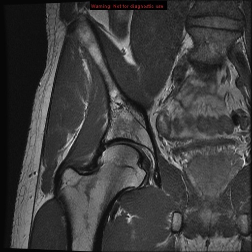
Hip MRI Anatomy
Detailed pelvic girdle anatomy, hip joint articulation, and surrounding musculature.
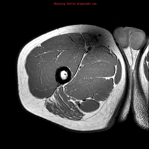
Thigh MRI Anatomy
Cross-sectional study of the quadriceps, hamstring compartments, and femoral vasculature.
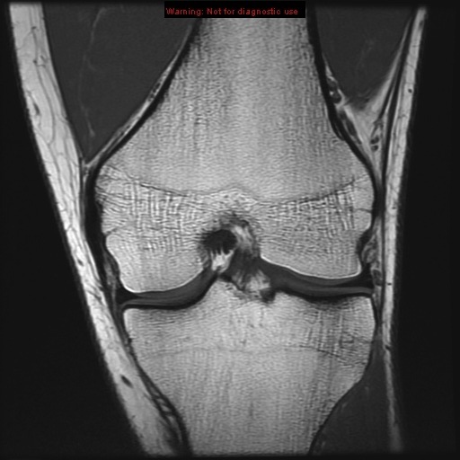
Knee MRI Anatomy
Cruciate ligaments, collateral ligaments, meniscus structures, and articular cartilage mapping.
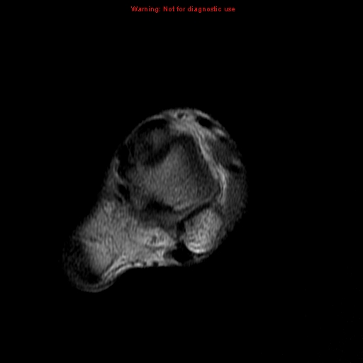
Lower Leg MRI Anatomy
Compartments of the calf, tibia/fibula structures, and muscular envelopes.
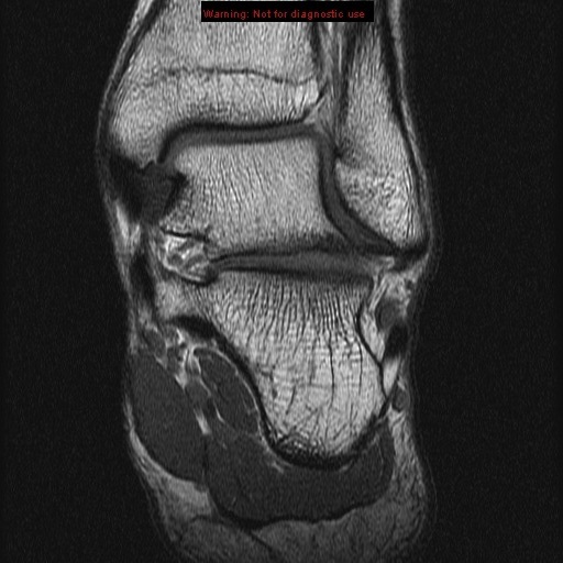
Ankle MRI Anatomy
Tarsal alignment, spring ligament, Achilles tendon, and complex ankle joint mapping.
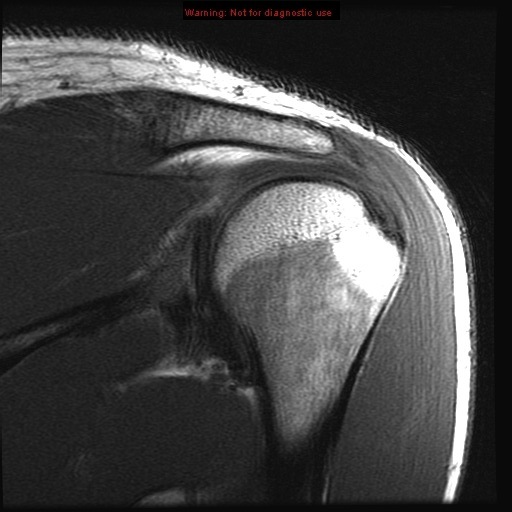
Shoulder MRI Anatomy
Rotator cuff tendons, glenoid labrum complexes, and biceps anchor pulleys.
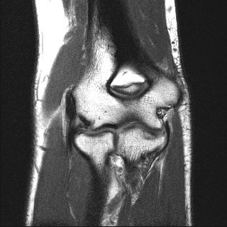
Elbow MRI Anatomy
Ulnar/radial collateral ligaments, bicipital insertions, and cubital tunnel neurography.
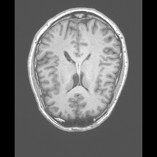
Brain MRI Anatomy
Interactive brain MRI mapping sagittal, coronal, and axial slices with lobes, ventricles, deep gray structures, and brainstem.
Clinical & FRCR Study Utility
These atlases map high-resolution MRI scans against standardized clinical terminologies. Toggle overlay modes for blind self-testing (hotspots only) or reference studies (lines + leader tags). Use scroll scrubbing to trace continuous anatomical runs.